


Vascular Doppler
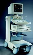 A Vascular Doppler is also referred to as a vascular ultrasound. Ultrasound testing uses high frequency sound waves to create an image of the area being studied. This is done with a transducer that is pressed against the skin. The transducer sends the sound waves, and records the image as the waves are reflected back. This image is instantly transmitted to a video monitor for the technician to see, and can be recorded on video.
For vascular doppler sonography, the technician will concentrate these sound waves on the patient's heart and blood vessels.
Vascular ultrasound provides pictures of the body's veins and arteries.
Why do I need Vascular Doppler procedure?
- help monitor the blood flow to organs and tissues throughout the body.
- locate and identify blockages (stenosis) and abnormalities like plaque or emboli and help plan for their effective treatment.
- detect blood clots (deep venous thrombosis (DVT) in the legs or arms.
- determine whether a patient is a good candidate for a procedure such as angioplasty.
- evaluate the success of procedures that graft or bypass blood vessels.
- determine if there is an enlarged artery (aneurysm).
- determine the source and severity of varicose veins.
Doppler ultrasound images can help the physician to see and evaluate:
- blockages to blood flow (such as clots).
- narrowing of vessels (which may be caused by plaque).
- tumors and congenital malformation.

Copyright ©1996-2012 Michigan Physicians Group. All Rights Reserved